Claremont | 08 9384 6855 Jandakot | 08 9414 1470
We are proud to deliver exceptional orthodontic care using state-of-the-art technology. Sophisticated equipment helps us more effectively diagnose orthodontic issues, understand your treatment needs better and deliver a customised treatment program for your unique circumstances.
We want to provide you the very best treatment outcome in the most biologically efficient way possible. Suresmile is redefining how orthodontic treatment is delivered, and has modernised orthodontics in Perth.
Orthodontic treatment has historically been a laborious process, often characterized by long treatment times, multiple visits, and varying results. Emerging customised technologies have sought to refine this process, and SureSmile is a prime example. Developed by OraMetrix, SureSmile combines 3D diagnostic imaging with computerized treatment planning and robotic wire bending to offer an innovative solution in orthodontics (Alford et al., 2011). SureSmile is a technologically advanced orthodontic system that provides orthodontists with a comprehensive 3D visualization platform to design and implement precise, patient-specific treatments. We have been mastering this technology for over 12 years, and have successfully treated thousands of patients in Perth over this period with this advanced treatment approach.
The SureSmile process begins with the collection of diagnostic data via advanced imaging technologies, typically cone-beam computed tomography (CBCT) or optical scanning. This data forms a detailed 3D model of the patient’s oral anatomy. The orthodontist then uses SureSmile’s treatment planning software to analyze the 3D model and formulate an individualized treatment plan, taking into account the final, desired positioning of the teeth.
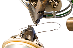
Once the treatment plan is established, it is sent to SureSmile’s robotic system, which precisely bends a shape-memory alloy archwire according to the planned movements. When applied, these custom archwires move teeth along the most efficient path to the pre-determined final position.
SureSmile’s digital treatment planning platform allows for greater precision in the formulation and implementation of treatment plans (Alford et al., 2011). The 3D model provides a comprehensive view of the patient’s oral anatomy, allowing the orthodontist to visualize and plan the exact desired position of each tooth. The robotically bent archwires enable more accurate force application, leading to the precise movement of teeth.
A study by Alford et al. (2011) found that patients treated with SureSmile technology completed their treatment 29.7% faster than those treated with conventional orthodontics. The efficiency of SureSmile arises from the use of custom, robotically bent wires, which require fewer adjustments and allow teeth to move along the most efficient path to their final positions.
With SureSmile’s precise movement planning, patients require fewer visits for wire changes or adjustments (Grauer et al., 2011). This not only reduces the burden on patients’ schedules but also minimizes discomfort and inconvenience associated with frequent orthodontic visits.
Greater precision, reduced treatment time, and fewer visits contribute to improved patient satisfaction. Patients have reported higher comfort levels, as the precise, smooth movement of teeth mitigates common discomforts associated with traditional orthodontic methods (Alford et al., 2011).
SureSmile technology represents a significant advancement in orthodontic treatment, offering greater precision, reduced treatment time, fewer visits, and increased patient satisfaction. Continued studies and technological advancements are anticipated to further optimize this method, improving the quality of care and treatment outcomes for orthodontic patients.
Suresmile has been shown to reduce treatment times by more than 30%, saving an average of 8 months in treatment.
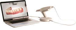
With the continuous evolution of digital technology in the dental field, intra-oral scanning has become a pivotal instrument in modern orthodontics. Over recent years, research has indicated that intra-oral scanners offer a high degree of precision, often comparable or even superior to traditional impression techniques. One of the major advantages of intra-oral scanning is the elimination of the need for physical impressions, which can be uncomfortable for patients, especially those with gag reflexes. Additionally, the digital impressions obtained can be instantly reviewed, modified, and shared, allowing for efficient communication between the orthodontist, dental technician, and other involved professionals.
Recent advancements in intra-oral scanning technology have enhanced not only the accuracy but also the speed of data acquisition. This swift capture of detailed 3D images aids in patient education, as they can immediately visualize their dental anatomy and the proposed treatment plans. Moreover, integration with other digital platforms, such as cone-beam computed tomography (CBCT) and digital treatment planning software, facilitates comprehensive orthodontic assessments and simulations. Such integrations pave the way for clearer treatment pathways, with potential reductions in treatment time and improved patient outcomes.
Furthermore, as the field continues to advance, we’re witnessing an increased emphasis on artificial intelligence and machine learning integration in intra-oral scanning systems. These augmentations can automate specific processes, like occlusion analysis and bracket placement simulations, streamlining the orthodontic workflow. Given the trajectory of research and technological progression, it’s evident that intra-oral scanning will remain at the forefront of orthodontic innovation, playing a pivotal role in enhancing both patient care and clinical efficiency.
Dr Siva was one of the first orthodontists to utilize intra-oral scanning technology, and conducted a randomized clinical trial in Boston over 10 years ago with a prototype of the 3M Lava True Definition scanner.
The end result is a superior-quality digital study model which we can use in our comprehensive treatment planning process. We can view your digital impressions onscreen from any angle and at any magnification to fully assess your clinical care requirements. We also use these digital models to custom-design appliances for patients choosing Invisalign, Suresmile and Incognito Hidden Braces.
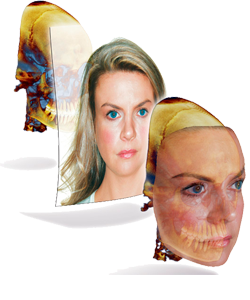
3D facial mapping technology has revolutionised the field of orthodontics, providing an incredibly detailed understanding of facial anatomy, growth patterns, and treatment outcomes. Unlike traditional 2D imaging methods, which only provide a limited view, 3D facial mapping generates a comprehensive representation of the craniofacial structures. It’s particularly beneficial when planning complex orthodontic procedures as it enables practitioners to visualise treatment effects on the entire facial profile, rather than just the teeth and jaws. With increased accuracy and predictability, orthodontists can formulate more precise treatment plans, reduce uncertainties, and potentially shorten treatment time.
The latest research highlights the integration of 3D facial mapping with other cutting-edge technologies like AI and machine learning to further advance treatment planning and monitoring. Predictive models can be developed using AI algorithms based on large data sets derived from 3D facial mapping. These models can forecast growth patterns, optimise treatment strategies, and even predict post-treatment outcomes. Moreover, these models allow for the customisation of treatments according to the patient’s unique facial structure and growth potential, enhancing the effectiveness of the interventions and resulting in better aesthetic and functional outcomes.
Looking forward, the continued advancements in 3D facial mapping technology promise even greater potential. The integration of 3D printing and CAD/CAM technology with 3D facial mapping can pave the way for creating personalised appliances, aligners and implants. This could revolutionise orthodontic practice by allowing more precise fabrication and fitting of orthodontic appliances. Furthermore, the use of cloud-based data storage can facilitate remote consultations and the global exchange of patient data, significantly broadening the reach and accessibility of specialist orthodontic care. Indeed, the potential of 3D facial mapping in orthodontics is vast, with opportunities for research, innovation, and practice improvement that can redefine the standards of care in orthodontics.
Dr Siva and his colleagues at Harvard University have applied this cutting edge technology for the simulation of clinical treatment plans for complex craniofacial case and the evaluation of surgical outcomes: Harvard University / Boston Children’s Hospital Cleft Surgery Research
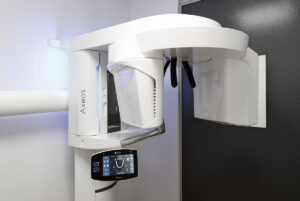
3D Cone Beam Computed Tomography (CBCT) technology has brought revolutionary advancements in the field of orthodontics, enabling clinicians to diagnose and treat patients more accurately and efficiently. One of the primary benefits of 3D CBCT is the ability to obtain a true three-dimensional image of the oral-maxillofacial complex. Unlike conventional 2D radiographic images, 3D CBCT technology provides a more comprehensive and accurate representation of patients’ anatomical structures, including details of the bones, teeth, and soft tissues. This increased precision helps in the meticulous planning of orthodontic treatments, particularly in complex cases such as impacted teeth, temporomandibular joint disorders, and craniofacial anomalies. It also greatly assists in the design and placement of dental implants and surgical guides.
In the realm of research, there are continuous developments in the optimization and application of CBCT technology. More recently, software improvements have allowed for more reliable measurements and better image quality, thereby reducing the need for higher radiation doses. AI-based algorithms are being integrated into CBCT systems, allowing for automatic detection and diagnosis of various conditions, which can enhance the diagnostic efficiency and accuracy of clinicians. Furthermore, CBCT technology is increasingly being integrated with digital orthodontics, including CAD/CAM and 3D printing technologies, which enables patient-specific treatment planning and fabrication of orthodontic appliances.
We use a highly sophisticated digital radiology system that minimizes radiation exposure for our patients. Our radiology machine was designed specifically for orthodontic treatment and automatically adjusts for each patient to obtain accurate images every time. The digital radiograph can be viewed immediately and downloaded directly into our imaging software.
Our digital radiology system is conveniently located on-site at Orthodontics on St Quentin and Orthodontics on Berrigan, which means you don’t need to travel to a third-party radiology clinic to obtain your images.
"The age of digital orthodontics is here."